


Facial Muscle Actions and Function: An Anatomy Guide for Acupuncturists
Understanding facial muscle actions helps acupuncturists connect visible expressions with the anatomy that produces them. In this American Acupuncture Council presentation, Michelle Gellis discusses facial muscle function in the context of facial acupuncture education. In addition, this companion article reviews the major muscle groups, their actions, and practical anatomical considerations for clinicians and students.
Watch the presentation: Open the video on YouTube.
Transcript: The original AAC listing advertises a downloadable transcript. The exact downloadable asset URL could not be independently retrieved during this revision. Restore the original “Click here to download the transcript” link from the existing WordPress media/link field before publication; do not replace it with an unverified URL.
Why facial muscle actions matter in facial acupuncture
Facial muscles do more than create expressions. For example, they help close the eyelids and lips, move the nostrils, support oral function, and coordinate movements used in speaking and eating. Moreover, unlike many skeletal muscles, numerous muscles of facial expression insert into skin or superficial connective tissue. As a result, their contraction changes the position of the overlying skin.
Most muscles of facial expression receive motor innervation from the facial nerve (cranial nerve VII). By contrast, the muscles of mastication, including the masseter and temporalis, are primarily supplied by the mandibular division of the trigeminal nerve (cranial nerve V3). Therefore, this distinction matters when assessing facial movement: not every muscle around the face belongs to the facial-expression group.
Key facial muscles and their primary actions
Forehead and eyebrow
- Frontal belly of the occipitofrontalis (frontalis): elevates the eyebrows and produces horizontal forehead lines.
- Corrugator supercilii: draws the eyebrows medially and downward, contributing to vertical glabellar lines.
- Procerus: pulls the medial brow downward and contributes to transverse lines over the bridge of the nose.
Periorbital region
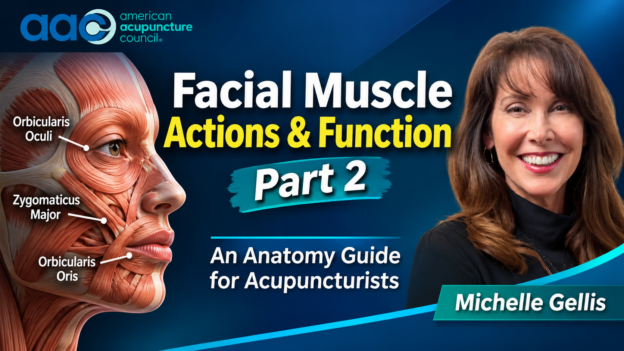
Orbicularis oculi encircles the eye. More specifically, its palpebral portion supports gentle eyelid closure, including blinking, while its orbital portion contributes to forceful closure. For this reason, the muscle is essential to normal eyelid function and should be distinguished from muscles that elevate the upper eyelid, which have different innervation and function.
Nose and upper lip
- Nasalis: its portions help compress or widen the nostrils.
- Levator labii superioris alaeque nasi: elevates the upper lip and can assist nostril dilation.
- Levator labii superioris: elevates the upper lip.
- Zygomaticus minor: elevates the upper lip and contributes to the nasolabial expression pattern.
Cheek and mouth
- Zygomaticus major: draws the corner of the mouth upward and laterally, contributing to smiling.
- Levator anguli oris: elevates the corner of the mouth.
- Risorius: draws the corner of the mouth laterally.
- Buccinator: compresses the cheek against the teeth and helps keep food between the dental surfaces during chewing. It also contributes to blowing and sucking.
- Orbicularis oris: closes and protrudes the lips; it is important for articulation and oral competence.
Lower lip, chin, and neck
- Depressor anguli oris: draws the corner of the mouth downward.
- Depressor labii inferioris: lowers and everts the lower lip.
- Mentalis: elevates and protrudes the lower lip and wrinkles the skin of the chin.
- Platysma: tenses superficial skin of the lower face and neck and can assist in depressing the lower lip and mandible.
Clinical interpretation: assess movement, not just appearance
Facial expressions usually arise from coordinated activity across several muscles rather than from one muscle acting alone. For example, a smile involves more than the zygomaticus major; the upper lip, eyelids, cheeks, and other perioral muscles contribute to the overall pattern. Consequently, functional assessment should compare movement at rest and during specific tasks, such as raising the eyebrows, closing the eyes gently and firmly, smiling, pursing the lips, showing the teeth, and puffing the cheeks.
For acupuncturists, anatomical knowledge can help organize observation, communication, and treatment planning. In practical terms, it can also help clinicians describe the region and tissue layer they intend to address. However, anatomy alone does not establish that needling a particular muscle will produce a specific cosmetic or functional result. Instead, treatment choices should reflect the patient’s presentation, practitioner training, scope of practice, and the limits of the available evidence.
Safety and professional considerations
Facial needling requires precise anatomical knowledge and appropriate training. Because the face contains variable superficial and deeper neurovascular structures, risk differs by location, depth, angle, and technique. Possible adverse events include pain, bleeding, bruising, and infection. Furthermore, deeper or poorly planned needling can create additional risk. For that reason, use clean-needle practices, obtain appropriate informed consent, apply conservative technique, and maintain a clear referral plan when findings fall outside your competence.
New or sudden facial weakness, asymmetry, difficulty closing an eye, speech changes, or other acute neurological symptoms require prompt medical assessment rather than an assumption that the problem is muscular or cosmetic. Likewise, persistent facial dysfunction warrants appropriate medical evaluation. Most importantly, acupuncture should never delay emergency assessment or indicated medical care.
Continue your facial acupuncture education
Explore these related American Acupuncture Council presentations:
- Treating the Face – Healing the Whole – Michelle Gellis
- Treating Neuromuscular Facial Conditions: A Multifaceted Approach
- Treating Cosmetic and Neuromuscular Facial Conditions with Facial Motor Points
- Facial Fascia: Appearance and Function
Conclusion
Knowing the actions and innervation of facial muscles gives acupuncturists a clearer framework for observing facial movement and discussing treatment goals. Ultimately, the most useful clinical approach combines anatomical precision with functional assessment, sound needling judgment, and appropriate referral. To explore these concepts further, watch Michelle Gellis’s presentation and use the transcript when AAC restores the original download link.
American Acupuncture Council: AAC supports the professional acupuncture community with educational resources and practice-focused information. Visit American Acupuncture Council to explore additional resources.
Educational disclaimer: This article is for professional education and general information. It does not provide individualized medical advice, establish a diagnosis, or replace appropriate medical evaluation. Practitioners are responsible for working within their training, licensure, and applicable laws and standards.
References
- Fakoya AO, Hohman MH, Westbrook KE, Varacallo M. Anatomy, Head and Neck: Facial Muscles. StatPearls [Internet]. Updated April 20, 2024. NCBI Bookshelf.
- Ringeisen AJ, et al. Facial Anatomy – Muscles. EyeWiki. Reviewed September 5, 2026. American Academy of Ophthalmology EyeWiki.
- Table 1. Facial Expression Muscles. StatPearls/NCBI Bookshelf. NCBI Bookshelf table.
- Cosmetic Acupuncture for Facial Aging: Anatomy, Mechanobiology, and Clinical Evidence—A Narrative Review. Journal of Cosmetic Dermatology. First published October 16, 2026. https://doi.org/10.1111/jocd.71245.